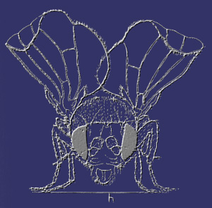
THUMMEL LAB RESEARCH SUMMARY
         

Overview:
Small lipophilic hormones, such as retinoic acid, steroids and thyroid hormone, exert profound effects on the growth, development, and physiology of higher organisms. Hormone action is mediated by members of the nuclear receptor superfamily that act as ligand-regulated transcription factors. Although we know a great deal about how nuclear receptors control target gene transcription, we have a relatively poor understanding of events downstream from the receptor – how effects on hormone-regulated gene expression result in the appropriate biological responses during development. Work in our lab is focused on defining these regulatory pathways using the fruit fly, Drosophila melanogaster, as a model system.

Much of our work focuses on the only known physiologically active steroid in insects, 20-hydroxyecdysone (referred to here as ecdysone), and its receptor EcR. Pulses of ecdysone act as a critical temporal signal, driving the animal forward in its life cycle by triggering each major developmental transition (Figure 1). An ecdysone pulse midway through embryonic development coordinates morphogenetic movements that establish the body plan of the first instar larva (Kozlova and Thummel, 2003). Ecdysone pulses during the first and second larval instars signal molting of the cuticle, defining the duration of each instar. A high titer ecdysone pulse at the end of the third instar triggers puparium formation and the onset of metamorphosis, initiating the prepupal stage of development. The larval midgut is destroyed in response to this late larval hormone pulse while the imaginal discs evert to form rudiments of the adult wings and legs. This is followed, ~10 hours later, by a low titer ecdysone pulse that signals the prepupal-pupal transition. In response to this prepupal ecdysone pulse, the adult head assumes its appropriate position by everting from inside the thorax, the legs and wings elongate, and the larval salivary glands are destroyed. The net result of these sequential ecdysone-triggered responses at the onset of metamorphosis is the transformation of a crawling larva into an immature adult fly during an ~14 hour interval. Studies with ecdysone-deficient mutants, as well as EcR mutants, have supported the model that ecdysone acts as a temporal cue, ensuring that the animal progresses to the next stage in the life cycle at the appropriate time (Thummel, 2001). A central question that arises from this observation is how the repeated systemic hormonal signal is refined into different responses at different times. A focus of work in our lab is aimed at defining the molecular mechanisms by which spatial and temporal specificity is conferred in this signal transduction pathway.


Figure 1. Pulses of ecdysone trigger each of the major developmental transitions in the life cycle. The developmental transitions are marked by dotted lines. The ecdysone titer profile is depicted as 20E equivalents in whole body homogenates (adapted from ref. Riddiford, 1993).
       
Ecdysone acts through regulatory cascades:
 
Ecdysone exerts its effects on development by regulating gene expression through its interaction with a heterodimer of two nuclear receptors: the EcR ecdysone receptor and the Drosophila RXR ortholog, USP. This hormone-receptor complex directly induces a number of primary-response target genes, including a small set of early genes that encode transcription factors. The early transcription factors, in turn, regulate large sets of downstream secondary-response late genes that direct the appropriate stage- and tissue-specific biological responses to the hormonal signal (Figure 2). A focus of work in our lab is to define these ecdysone-triggered regulatory cascades.
Figure 2. Ecdysone-triggered regulatory cascades. Classic studies of the puffing patterns of the giant larval salivary gland polytene chromosomes defined a regulatory cascade for ecdysone action (Ashburner et al., 1974). The hormone/receptor complex directly induces early regulatory genes that, in turn, control large sets of late effector genes, directing the appropriate biological responses to the hormone.
         
Toward this goal, we have used microarrays to identify genes regulated by 20E alone or 20E in the presence of the protein synthesis inhibitor cycloheximide (Beckstead et al., 2005). We have also examined the effects of disrupting EcR function on the global patterns of gene expression at the onset of metamorphosis, and used this data to refine our lists of 20E-regulated genes. This study identified many new genes that are part of the 20E/EcR regulatory cascade and defined roles for EcR in the regulation of stress, immunity, and metabolism at the onset of metamorphosis. We are building off this foundation by performing reverse genetic studies of new transcription factor-encoding genes that are directly regulated by the 20E/EcR complex.
         
Programmed Cell Death:
Ecdysone directs two main biological programs during metamorphosis – the massive death of obsolete larval tissues and their replacement by new adult tissues and structures (see our movies of this). A central focus of our research is aimed at defining the molecular mechanisms by which the hormone controls these divergent developmental pathways.
 
Figure 3. Ecdysone triggers the stage-specific cell death of larval tissues at the onset of metamorphosis. The late larval ecdysone pulse (20E box on left) triggers larval midgut cell death, while the salivary glands survive until the prepupal pulse (20E box on right), dying immediately after adult head eversion.
         

Early work in our lab showed that the destruction of larval tissues is a stage-specific steroid-triggered response that has hallmark features of programmed cell death (Figure 3), including DNA fragmentation and caspase activation. We also found that this response is foreshadowed by the coordinate induction of two key death activators, reaper and hid (Jiang et al., 1997) (Figure 4). Our functional studies indicate that these genes act together, in a partially redundant manner, to direct the death response, and that premature activation of this pathway is prevented by the DIAP1 death inhibitor (Yin and Thummel, 2004). We have also shown that reaper is directly regulated by EcR through an essential response element in its promoter, and that the BR-C and E74A ecdysone-inducible transcription factors are required for proper reaper and hid expression and salivary gland cell death (Jiang et al., 2000) (Figure 4). Taken together, this work provides the first description of a steroid-triggered gene cascade that directs a programmed cell death response, establishing a framework for understanding how similar pathways might be controlled in other organisms.

We are currently using genetic screens to expand our understanding of steroid-triggered salivary gland cell death. By using a salivary gland-specific GFP marker, we can follow the fate of this tissue in living animals (see our movies of this). We are screening for mutations that lead to appropriate progression through the prepupal-to-pupal transition – with relatively normal head eversion and leg elongation – but persistent larval glands. Pilot screens using P element mutations resulted in the identification of a number of genes in this pathway, including E74 and hid, validating this approach and providing us with several genes that we are currently characterizing. We are also expanding this screen, using EMS to generate mutations on the third chromosome that result in larval salivary gland death defects. To date, this effort has resulted in the recovery of seven multiallelic complementation groups that we are currently mapping to specific genes. We anticipate that the mechanisms of ecdysone-triggered cell death will provide a valuable paradigm for understanding how steroids control stage- and tissue-specific biological responses during development, as well as provide new insights into the regulation of programmed cell death.
 
Adult tissue development:

One of the main impediments to studying metamorphosis is the opaque pupal cuticle that prevents visualization of the developmental changes occurring within. We have overcome this problem by expressing GFP in specific tissues and making time-lapse movies to follow the fate of those tissues in living animals (see our movies of metamorphosis). These movies reveal, for the first time, the dynamics and coordination of morphogenetic movements that could only be inferred from earlier studies, as well as responses not previously described (Ward et al., 2003b). They also provide a rapid and simple means of discovering the origin of specific mutant phenotypes and a foundation for genetic screens, as described above.

Many studies have focused on how imaginal discs undergo proliferation and patterning during larval development, such that every cell in the mature disc is fated to become a specific part of the adult fly. In contrast, much less is known about how these discs undergo terminal differentiation, a process that is absolutely dependent on the sequential pulses of ecdysone that initiate metamorphosis. We would like to understand the molecular mechanisms by which ecdysone directs this transformation from a single cell layer disc epithelium to its corresponding adult structure. Mutations in EcR, BR-C, E74, ßFTZ-F1, crooked-legs, and other ecdysone-regulated genes result in characteristic leg malformations that can be easily scored in pupal stages (Broadus et al., 1999; D'Avino and Thummel, 1998). Past efforts in our lab have used genetic screens to approach this problem, identifying a key role for the Rho GTPase signal transduction pathway in mediating the effects of ecdysone on leg morphogenesis (Gates and Thummel, 2000; Ward et al., 2003a).

       
Nuclear Receptor Regulation & Function:

A major effort underway in the lab is to define roles for nuclear receptors in development, growth, and metabolism. The Drosophila genome encodes 18 canonical nuclear receptors, with representatives of all the major vertebrate classes, providing the smallest complete set of receptors known in any genetic model system (King-Jones and Thummel, 2005) (Figure 5). We have determined the temporal profiles of expression for all detectable nuclear receptor genes during the major ecdysone-triggered transitions in the life cycle (Sullivan and Thummel, 2003). We are continuing our reverse genetic characterization of this gene family, focusing on nuclear receptors that have close mammalian homologs, in an effort to understand their functions during development (for a recent example, see our studies of DHR4 in ref. King-Jones et al., 2005). We analyze the phenotypes associated with both loss-of-function and gain-of-function mutations as a means of determining gene function. Using heat-inducible expression of dsRNA for RNAi provides us with the temporal control that is needed to study the later functions of genes that are required for early development (Lam and Thummel, 2000).

Figure 5. Phylogeny of human and fly nuclear receptors. Drosophila encodes only 18 nuclear receptors (red) in comparison to the 48 receptors encoded by the human genome (black) (Robinson-Rechavi et al., 2003). In spite of this small number, the fly nuclear receptors represent all major subclasses of human receptors and include orthologs of key human receptors, providing a good model for studying nuclear receptor regulation and function.

By raising antibodies against the encoded proteins we can determine their spatial patterns of expression as well as their binding sites in the giant polytene chromosomes, identifying potential direct regulatory targets. We use microarrays to determine the effects of loss-of-functionand gain-of-function mutations on gene expression, and classify these targets into functional groups by bioinformatics. Most recently, we have used metabolic profiling to determine effects of nuclear receptor mutations on specific metabolic pathways, linking these functions to key target genes identified in our microarray studies as well as specific developmental defects. Taken together, our goal is to use the fly as a model system for understanding how nuclear receptors regulate growth, metabolism, and development, with a focus on understanding the crosstalk between critical metabolic checkpoints and developmental transitions. Current work is focused on dERR, DHR38, DHR78, DHR96, and dHNF4 (Figure 5). We also use transgenic animals that carry the GAL4 DNA binding domain fused to the ligand binding domains (LBDs) of nuclear receptors as a means of determining when and where hormones or critical co-factors are present in the animal (Kozlova and Thummel, 2002). In collaboration with Henry Krause’s lab (Univ. of Toronto) we have established multiple transformant lines that express GAL4-LBD fusions for all 18 fly receptors, and we have completed the characterization of their activation patterns during embryogenesis and the onset of metamorphosis (Palanker et al., 2006). These studies have identified novel agonists for some nuclear receptors, preferred sites of LBD activation in tissues associated with lipid metabolism, and dynamic changes in activation indicative of novel hormones or cofactors that regulate receptor function. Taken together, these studies should clarify the roles of nuclear receptors in development, growth and metabolism, and provide new insights into the regulation and function of their vertebrate orthologs.

         
References (click on references below from Thummel lab to download PDF file):

Ashburner, M., Chihara, C., Meltzer, P. and Richards, G. (1974). Temporal control of puffing activity in polytene chromosomes. Cold Spring Harb Symp Quant Biol 38, 655-62.

Beckstead, R. B., Lam, G. and Thummel, C. S. (2005). The genomic response to 20-hydroxyecdysone at the onset of Drosophila metamorphosis. Genome Biol 6, R99.

Broadus, J., McCabe, J. R., Endrizzi, B., Thummel, C. S. and Woodard, C. T. (1999). The Drosophila bFTZ-F1 orphan nuclear receptor provides competence for stage-specific responses to the steroid hormone ecdysone. Mol Cell 3, 143-9.

D'Avino, P. P. and Thummel, C. S. (1998). crooked legs encodes a family of zinc finger proteins required for leg morphogenesis and ecdysone-regulated gene expression during Drosophila metamorphosis. Development 125, 1733-45.

Gates, J. and Thummel, C. S. (2000). An enhancer trap screen for ecdysone-inducible genes required for Drosophila adult leg morphogenesis. Genetics 156, 1765-76.

Jiang, C., Baehrecke, E. H. and Thummel, C. S. (1997). Steroid regulated programmed cell death during Drosophila metamorphosis. Development 124, 4673-83.

Jiang, C., Lamblin, A. F., Steller, H. and Thummel, C. S. (2000). A steroid-triggered transcriptional hierarchy controls salivary gland cell death during Drosophila metamorphosis. Mol Cell 5, 445-55.

King-Jones, K., Charles, J. P., Lam, G. and Thummel, C. S. (2005). The ecdysone-induced DHR4 orphan nuclear receptor coordinates growth and maturation in Drosophila. Cell 121, 773-84.

King-Jones, K. and Thummel, C. S. (2005). Nuclear receptors - a perspective from Drosophila. Nat Rev Genet 6, 311-23.

Kozlova, T. and Thummel, C. S. (2002). Spatial patterns of ecdysteroid receptor activation during the onset of Drosophila metamorphosis. Development 129, 1739-50.

Kozlova, T. and Thummel, C. S. (2003). Essential roles for ecdysone signaling during Drosophila mid-embryonic development. Science 301, 1911-4.

Lam, G. and Thummel, C. S. (2000). Inducible expression of double-stranded RNA directs specific genetic interference in Drosophila. Curr Biol 10, 957-63.

Palanker, L., Sampson, H., Necakov, A., Ni, R., Hu, C., Thummel, C. S. and Krause, H. (2006). Dynamic regulation of Drosophila nuclear receptor activity in vivo. Development, in revision.

Riddiford, L. M. (1993). Hormones and Drosophila development. In The Development of Drosophila melanogaster, vol. 2 (ed. M. Bate and A. Martinez-Arias), pp. 899-939. Cold Spring Harbor: Cold Spring Harbor Laboratory Press.

Robinson-Rechavi, M., Escriva Garcia, H. and Laudet, V. (2003). The nuclear receptor superfamily. J Cell Sci 116, 585-6.

Sullivan, A. A. and Thummel, C. S. (2003). Temporal profiles of nuclear receptor gene expression reveal coordinate transcriptional responses during Drosophila development. Mol Endocrinol 17, 2125-37.

Thummel, C. S. (2001). Molecular mechanisms of developmental timing in C. elegans and Drosophila. Dev Cell 1, 453-65.

Ward, R. E., Evans, J. and Thummel, C. S. (2003a). Genetic modifier screens in Drosophila demonstrate a role for Rho1 signaling in ecdysone-triggered imaginal disc morphogenesis. Genetics 165, 1397-415.

Ward, R. E., Reid, P., Bashirullah, A., D'Avino, P. P. and Thummel, C. S. (2003b). GFP in living animals reveals dynamic developmental responses to ecdysone during Drosophila metamorphosis. Dev Biol 256, 389-402.

Yin, V. P. and Thummel, C. S. (2004). A balance between the diap1 death inhibitor and reaper and hid death inducers controls steroid-triggered cell death in Drosophila. Proc Natl Acad Sci U S A 101, 8022-7.